Using a new imaging technique, researchers have confirmed what
scientists have always thought to be true: the structural connections in
the brain are unique to each individual person.
The Carnegie Mellon University-led team used diffusion MRI to map
the brain's structural connections and found each person's connections
are so unique they could identify a person based on this brain
"fingerprint" with nearly perfect accuracy. Published in PLOS Computational Biology,
the results also show the brain's that distinctiveness changes over
time, which could help researchers determine how factors such as
disease, the environment and different experiences impact the brain.
The new, non-invasive diffusion MRI approach captures the brain's
connections at a much closer level than ever before. For example,
conventional approaches obtain a single estimate of the integrity of a
single structural connection, or a white matter fiber. The new technique
measures the integrity along each segment of the brain's biological
wires, making it much more sensitive to unique patterns.
"The most exciting part is that we can apply this new method to
existing data and reveal new information that is already sitting there
unexplored. The higher specificity allows us to reliably study how
genetic and environmental factors shape the human brain over time,
thereby opening a gate to understand how the human brain functions or
dysfunctions," said Fang-Cheng (Frank) Yeh, the study's first author and
assistant professor of neurological surgery at the University of
Pittsburgh. Yeh completed the research while at CMU as a postdoctoral
fellow in psychology.
For the study, the researchers used diffusion MRI to measure the
local connectome of 699 brains from five data sets. The local connectome
is the point-by-point connections along all of the white matter
pathways in the brain, as opposed to the connections between brain
regions. To create a fingerprint, they took the data from the diffusion
MRI and reconstructed it to calculate the distribution of water
diffusion along the cerebral white matter's fibers.
The measurements revealed that the local connectome is highly unique
to an individual and can be used as a personal marker for human
identity. To test the uniqueness, the team ran more than 17,000
identification tests. With nearly 100 percent accuracy, they were able
to tell whether two local connectomes, or brain "fingerprints," came
from the same person or not.
Additionally, they discovered that identical twins only share about
12 percent of structural connectivity patterns and the brain's unique
local connectome is sculpted over time, changing at an average rate of
13 percent every 100 days.
"This confirms something that we've always assumed in neuroscience
-- that connectivity patterns in your brain are unique to you," said
CMU's Timothy Verstynen, assistant professor of psychology. "This means
that many of your life experiences are somehow reflected in the
connectivity of your brain. Thus we can start to look at how shared
experiences, for example poverty or people who have the same
patholoigical disease, are reflected in your brain connections, opening
the door for potential new medical biomarkers for certain health
concerns."
In addition to Yeh and Verstynen, the research team included CMU's
Aarti Singh and Barnabas Poczos, the U.S. Army Research Laboratory's
Jean M. Vettel, the University of California, Santa Barbara's Scott T.
Grafton, the University of Pittsburgh's Kirk I. Erickson and Wen-Yih I.
Tseng of the National Taiwan University.
The Army Research Laboratory funded this research.
Developing a way to fingerprint the brain is one of the many brain
research breakthroughs to happen at Carnegie Mellon. CMU has created
some of the first cognitive tutors, helped to develop the
Jeopardy-winning Watson, founded a groundbreaking doctoral program in
neural computation, and is the birthplace of artificial intelligence and
cognitive psychology. Building on its strengths in biology, computer
science, psychology, statistics and engineering, CMU launched BrainHub,
an initiative that focuses on how the structure and activity of the
brain give rise to complex behaviors.
https://www.eurekalert.org/pub_releases/2016-11/cmu-rdw111416.php
Showing posts with label BRAIN. Show all posts
11/30/16
5/26/16
Science of Awakening
Introduction.
Recent advances in brain research using brain imaging techniques such
as SPECT, fMRI and EEG have indicated that the human brain is already
hard wired for enlightenment. It seems that the brain, over millions of
years of evolution, has been prepared for the experience of unity
with Cosmos or oneness with God.
Andrew Newberg, professor of nuclear medicine at the University of Pennsylvania,
is author of the acclaimed book ‘Why God Won’t Go Away’. In an attempt
to bridge science and spirit Newberg studied eight Tibetan Buddhist
practitioners during meditation using SPECT scan. The images he captured
showed that the brain’s prefrontal cortex during deep meditation lit up
in a red color indicating an increase in blood flow and neural activity
in that area. At the same time, surprisingly, the upper rear part of
the brain called the parietal area turned a dark blue shade indicating a
sudden drop of brain activity in that area which Newberg calls the
Orientation Association Area (OAA).
Newberg theorizes that when the meditator withdraws from the outside world, sensory input to the OAA is blocked and the neural activity in that area is shut down. At the same time due to the intense concentration (on a mantra, on God or guru) the prefrontal cortex or the Attention Association Area (AAA) is strongly activated and will now assume the role as the brain’s new experiential center.
The
OAA is the area which gives us the ability to orient ourselves in space
and time and which gives our bodies a sense of physical limits and the
self a sense of separateness from the rest of the universe. When the
OAA is deactivated the physical limits of the body and the sense of
separateness disappears. The brain can no longer create a boundary
between self and the outside world, or locate itself in physical
reality. As a result, Newberg says, the brain has no choice but to
perceive that self as endless, interwoven with everyone and everything. This is the state Newberg calls Absolute Unitary Being. We prefer to call it the Oneness State.
Newberg’s
research suggests that the process of awakening is not only due to
psychological change or a change in philosophy and values. No, it is
primarily due to a fundamental change in brain function with a shift in
brain dominance from the parietal (OAA) to the prefrontal (AAA) area.
When the over-activity in the OAA is decreased and the under-activity
in the AAA is increased, there is a shift of the brain’s command center
and the individual wakes up to a higher level of consciousness and
to a new reality which seems to be even more real than the old one.
Richard Davidson. Some of Newberg’s findings have been corroborated by neuroscientist Richard Davidson, University of Wisconsin. Davidson collaborated with Tibet’s Dalai Lama who sent eight of his most accomplished meditators to Davidson’s laboratory for a scientific study.
Using
both EEG and fMRI scans, Davidson studied the monks during deep
meditation and found very high activity in the prefrontal cortex -
especially on the left side which has to do with feelings of joy,
happiness and compassion. The EEG recordings during deep meditation
showed extremely powerful Gamma waves in that same area of the brain.Since there were no detailed descriptions of the monks’ levels of spiritual development in the above studies we have no idea whether any of them were in a permanent awakened state.
http://www.newbrainnewworld.com/?Science_of_Awakening
4/28/16
New Evidence Points to Personal Brain Signatures
Everyone's brain is
different. Until recently neuroscience has tended to gloss this over by
averaging results from many brain scans in trying to elicit general
truths about how the organ works. But in a major development within the
field researchers have begun documenting how brain activity differs
between individuals. Such differences had been largely thought of as
transient and uninteresting but studies are starting to show that they
are innate properties of people's brains, and that knowing them better
might ultimately help treat neurological disorders.
The latest study, published April 8 in Science, found that the brain activity of individuals who were just biding their time in a brain scanner contained enough information to predict how their brains would function during a range of ordinary activities. The researchers used these at-rest signatures to predict which regions would light up—which groups of brain cells would switch on—during gambling, reading and other tasks they were asked to perform in the scanner. The technique might be used one day to assess whether certain areas of the brains of people who are paralyzed or in a comatose state are still functional, the authors say.
The study capitalizes on a relatively new method of brain imaging that looks at what is going on when a person essentially does nothing. The technique stems from the mid-1990s work of biomedical engineer Bharat Biswal, now at New Jersey Institute of Technology. Biswal noticed that scans he had taken while participants were resting in a functional magnetic resonance imaging (fMRI) scanner displayed orderly, low-frequency oscillations. He had been looking for ways to remove background noise from fMRI signals but quickly realized these oscillations were not noise. His work paved the way for a new approach known as resting-state fMRI.
This type of scan, it turns out, reveals a lot about a particular brain. It analyzes the commonplace slow fluctuations of neural signaling, which form networks of brain cells that fluctuate in synchrony—and these networks often resemble those the brain engages when it is actively doing something. “We've known for awhile that the brain networks we pull out of resting-state data look similar to the maps we get from task-induced activity,” says neuroscience doctoral student Emily Finn of Yale University. Finn and her colleagues published a study last October showing that brain networks contain enough information to identify individuals with up to 99 percent accuracy. “This study takes things a step further,” Finn says.
The team behind the new study, led by neuroscientists Ido Tavor and Saad Jbabdi of the University of Oxford, used data collected by the Human Connectome Project (HCP)—a National Institutes of Health collaboration that is trying to map the wiring of the human brain and is led by Washington University in Saint Louis, the University of Minnesota and Oxford University. The team obtained data for 98 healthy young adults, including scans taken while the participants performed tasks involving memory, motor functions, decision-making (gambling), language (reading) and others as well as just resting. They analyzed the relationships between participants' resting-state brain activity and the oscillations that emerged while they were engaged in various undertakings. They then tried to predict brain activity profiles for a given participant on each of the tasks, using only the individual’s resting-state scan. The predictions matched the brain activity of that person more closely than any of the other participants' scans. “We extract a set of images that highlight brain areas that fluctuate together during this mind-wandering state,” Jbabdi explains. “Our study shows that these co-fluctuations contain enough information to predict how the brain behaves when it is actually doing something explicit.”
These are only first steps. What other information might be contained in the resting-state scans, and how the relationship between resting and active states might change under some circumstances, remain open questions. “It will be interesting to see if and how this mapping relates to actual performance on the tasks,” Finn says. “And how it changes with factors like age or neuropsychiatric illness.”
Tavor says his group was impelled to do this study by a common problem neuroscientists face. For many studies, researchers need to know exactly which brain areas are chugging along during certain tasks—so (for instance) they can see what happens when they block or enhance that activity. The new technique could allow researchers to predict where these regions are without having to conduct a separate scan for each of the tasks, saving time and money. “It's a very practical result,” Finn says. “Resting-state could eventually serve as a “one-size-fits-all” scan from which we can glean a lot of information about someone, without actually having them sit though multiple task sessions in the scanner,” she adds.
One of the next endeavors in this research is to determine whether these findings hold not just for the healthy participants used in this study but for patients with various illnesses. “We're looking at brain tumor patients before surgery,” Tavor says. Knowing what parts of the brain are responsible for sensitive functions, like language, can be crucial information to a neurosurgeon, and tumors can cause shifts in where functions are performed in the brain. “If we can predict this shift, it could affect the surgeon's strategy of where to enter to remove the tumor,” Tavor explains.
Biswal is also interested in medical implications. “In clinical cases, if there's a difference in performance, compared to healthy controls, would the resting-state still predict patients' performance?” he asks. “Or has something mechanistic happened that means the prediction won't be as good, and might this tell us something about the underlying mechanism of the disease?” Using the technique for diagnostic applications might enable researchers to measure disease severity by examining the accuracy of predictions for brain functions known to be affected by a particular disease.
Whatever the eventual outcome, this work adds to a body of evidence suggesting the resting brain is anything but. “During this so-called resting-state, the brain is not really resting,” Tavor says. “It does everything, all the time.”
http://www.scientificamerican.com/section/news/new-evidence-points-to-personal-brain-signatures1/
The latest study, published April 8 in Science, found that the brain activity of individuals who were just biding their time in a brain scanner contained enough information to predict how their brains would function during a range of ordinary activities. The researchers used these at-rest signatures to predict which regions would light up—which groups of brain cells would switch on—during gambling, reading and other tasks they were asked to perform in the scanner. The technique might be used one day to assess whether certain areas of the brains of people who are paralyzed or in a comatose state are still functional, the authors say.
The study capitalizes on a relatively new method of brain imaging that looks at what is going on when a person essentially does nothing. The technique stems from the mid-1990s work of biomedical engineer Bharat Biswal, now at New Jersey Institute of Technology. Biswal noticed that scans he had taken while participants were resting in a functional magnetic resonance imaging (fMRI) scanner displayed orderly, low-frequency oscillations. He had been looking for ways to remove background noise from fMRI signals but quickly realized these oscillations were not noise. His work paved the way for a new approach known as resting-state fMRI.
This type of scan, it turns out, reveals a lot about a particular brain. It analyzes the commonplace slow fluctuations of neural signaling, which form networks of brain cells that fluctuate in synchrony—and these networks often resemble those the brain engages when it is actively doing something. “We've known for awhile that the brain networks we pull out of resting-state data look similar to the maps we get from task-induced activity,” says neuroscience doctoral student Emily Finn of Yale University. Finn and her colleagues published a study last October showing that brain networks contain enough information to identify individuals with up to 99 percent accuracy. “This study takes things a step further,” Finn says.
The team behind the new study, led by neuroscientists Ido Tavor and Saad Jbabdi of the University of Oxford, used data collected by the Human Connectome Project (HCP)—a National Institutes of Health collaboration that is trying to map the wiring of the human brain and is led by Washington University in Saint Louis, the University of Minnesota and Oxford University. The team obtained data for 98 healthy young adults, including scans taken while the participants performed tasks involving memory, motor functions, decision-making (gambling), language (reading) and others as well as just resting. They analyzed the relationships between participants' resting-state brain activity and the oscillations that emerged while they were engaged in various undertakings. They then tried to predict brain activity profiles for a given participant on each of the tasks, using only the individual’s resting-state scan. The predictions matched the brain activity of that person more closely than any of the other participants' scans. “We extract a set of images that highlight brain areas that fluctuate together during this mind-wandering state,” Jbabdi explains. “Our study shows that these co-fluctuations contain enough information to predict how the brain behaves when it is actually doing something explicit.”
These are only first steps. What other information might be contained in the resting-state scans, and how the relationship between resting and active states might change under some circumstances, remain open questions. “It will be interesting to see if and how this mapping relates to actual performance on the tasks,” Finn says. “And how it changes with factors like age or neuropsychiatric illness.”
Tavor says his group was impelled to do this study by a common problem neuroscientists face. For many studies, researchers need to know exactly which brain areas are chugging along during certain tasks—so (for instance) they can see what happens when they block or enhance that activity. The new technique could allow researchers to predict where these regions are without having to conduct a separate scan for each of the tasks, saving time and money. “It's a very practical result,” Finn says. “Resting-state could eventually serve as a “one-size-fits-all” scan from which we can glean a lot of information about someone, without actually having them sit though multiple task sessions in the scanner,” she adds.
One of the next endeavors in this research is to determine whether these findings hold not just for the healthy participants used in this study but for patients with various illnesses. “We're looking at brain tumor patients before surgery,” Tavor says. Knowing what parts of the brain are responsible for sensitive functions, like language, can be crucial information to a neurosurgeon, and tumors can cause shifts in where functions are performed in the brain. “If we can predict this shift, it could affect the surgeon's strategy of where to enter to remove the tumor,” Tavor explains.
Biswal is also interested in medical implications. “In clinical cases, if there's a difference in performance, compared to healthy controls, would the resting-state still predict patients' performance?” he asks. “Or has something mechanistic happened that means the prediction won't be as good, and might this tell us something about the underlying mechanism of the disease?” Using the technique for diagnostic applications might enable researchers to measure disease severity by examining the accuracy of predictions for brain functions known to be affected by a particular disease.
Whatever the eventual outcome, this work adds to a body of evidence suggesting the resting brain is anything but. “During this so-called resting-state, the brain is not really resting,” Tavor says. “It does everything, all the time.”
http://www.scientificamerican.com/section/news/new-evidence-points-to-personal-brain-signatures1/
4/3/16
Brain scan may reveal if you are a true altruist or driven by self-interest
The reason why we help others at a cost to ourselves
has long presented a puzzle for scientists. Why do some of us do it
more than others? And are we doing it because we are truly moved by the
suffering of others or simply because we feel we ought to return a favour
or even get something in return? Looking at behaviour alone, it can be
hard to tell. Both empathy and the principle of reciprocity - giving to
return a favour or expecting others to do so - are proposed explanations
for altruism which have been impossible to separate until now.
Using functional magnetic resonance imaging (fMRI), which measures blood flow changes in the brain, a new study
suggests that specific differences in connectivity between brain
regions can predict whether someone is an empathy-driven altruist, a
reciprocity-driven altruist - or just selfish.
In the experiment, 34 female participants were divided into two groups.
Those in the "empathy" group witnessed an actor receive painful electric
shocks - and received shocks themselves (so they knew it hurt). In the
"reciprocity" group, participants were paired up with actors who kindly
paid money so the participant received fewer shocks (although both
groups received the same number of shocks overall).
Next, their brains were scanned. During the scanning, participants were
asked to split a sum of money between themselves and another person. For
the empathy group, the other person receiving the money was sometimes
the partner they saw shocked. In the reciprocity group, the person was
sometimes the partner who paid for the participant to receive fewer
shocks. At other times, participants were simply asked to split the cash
between themselves and a neutral person who neither received shocks nor
did anything nice. The researchers could therefore divide the
participants into those empathy-driven altruists and reciprocity-driven
altruists based on the first part of the experiment. They could also use
the way participants split the money in the second part to identify
selfish individuals among these participants.
Unsurprisingly, the initial analysis showed that participants gave, on
average, larger sums of money to the empathy and reciprocity partners
than to the neutral partner - and that both groups were equally
generous. Those that most regularly chose splits involving more money
for themselves than the other were classified as "selfish". But this was
just the starting point. The researchers used a complex and
sophisticated follow-up to gain deeper insight.
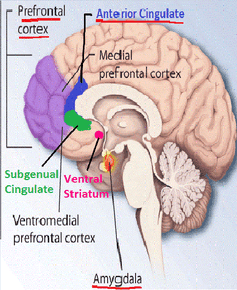
By looking at the timing of activity in the anterior cingulate cortex
(known for a host of functions from pain and conflict to learning), the
anterior insula cortex (associated with arousal and emotion) and the
ventral striatum (associated with rewards and learning), the researchers
created models of how information was passed between these areas. Then a
computer algorithm tried to guess, based on these models, whether an
individual's altruistic decision had been motivated by empathy or by
reciprocity. The high accuracy of these guesses at 77% shows the two
groups of participants had brain activity patterns that differed enough
to classify.
In empathy-driven altruism, the anterior insula (emotion and arousal)
and ventral striatum (rewards) showed a lower than average connectivity,
while reciprocity-driven altruism showed increased connectivity between
these regions. Connectivity in this sense can be imagined as how much
one area is "talking to" another. Although the functions of these areas
are broadly known, the meaning of changes in connectivity is still
difficult to interpret.
By looking at the timing of activity in the anterior cingulate cortex
(known for a host of functions from pain and conflict to learning), the
anterior insula cortex (associated with arousal and emotion) and the
ventral striatum (associated with rewards and learning), the researchers
created models of how information was passed between these areas. Then a
computer algorithm tried to guess, based on these models, whether an
individual's altruistic decision had been motivated by empathy or by
reciprocity. The high accuracy of these guesses at 77% shows the two
groups of participants had brain activity patterns that differed enough
to classify.
In empathy-driven altruism, the anterior insula (emotion and arousal)
and ventral striatum (rewards) showed a lower than average connectivity,
while reciprocity-driven altruism showed increased connectivity between
these regions. Connectivity in this sense can be imagined as how much
one area is "talking to" another. Although the functions of these areas
are broadly known, the meaning of changes in connectivity is still
difficult to interpret.
Can we learn to be more altruistic?
When it comes to implications, the differences between primarily selfish or primarily altruistic participants may be the most important finding. Inducing empathy, by seeing someone shocked, increased giving and associated neural connectivity for selfish individuals - they were more generous to the shocked partners than to the neutral person. The altruistic people, however, shared just as much with the neutral person as the shocked partner. The opposite was true for the reciprocity effect: increased giving to the partner who paid to prevent their shocks was seen in altruistic but not selfish participants.
One could speculate that this implies that altruistic participants are already giving because of empathic motivation, so increasing empathy makes no difference - they are at their "empathy capacity". Similarly, selfish participants may already be acting due to motivations more likely to benefit themselves too, such as reciprocity.
Research on altruism regularly concludes that people have an empathetic motivation but this paper suggests potential for future studies to check whether this is the case for each individual participant. The authors also open doors to more specific measures and targets for further research on reciprocity and empathy.
When it comes to implications, the differences between primarily selfish or primarily altruistic participants may be the most important finding. Inducing empathy, by seeing someone shocked, increased giving and associated neural connectivity for selfish individuals - they were more generous to the shocked partners than to the neutral person. The altruistic people, however, shared just as much with the neutral person as the shocked partner. The opposite was true for the reciprocity effect: increased giving to the partner who paid to prevent their shocks was seen in altruistic but not selfish participants.
One could speculate that this implies that altruistic participants are already giving because of empathic motivation, so increasing empathy makes no difference - they are at their "empathy capacity". Similarly, selfish participants may already be acting due to motivations more likely to benefit themselves too, such as reciprocity.
Research on altruism regularly concludes that people have an empathetic motivation but this paper suggests potential for future studies to check whether this is the case for each individual participant. The authors also open doors to more specific measures and targets for further research on reciprocity and empathy.
The paper shows the importance of analysing subtle differences in brain communication rather than overall activity.
Looking at different brain regions working together, rather than in
isolation, can identify previously elusive psychological concepts, such
as underlying motivations.
Future research is needed on whether these increases in altruism and neural connectivity could last, perhaps with ongoing training. For example, if the techniques used to induce empathy in the study could be employed in some sort of treatment for antisocial behaviour.
However, charities can already make the most of the current findings. They suggest empathy-inducing appeals may be most effective for new supporters, who are not yet "altruistic enough" to donate. Existing supporters, who are already altruistic, may respond more to receiving a token gift they feel they can reciprocate by increasing their donations. The effectiveness of these techniques, already used by many charities, may be explained by the findings. But with limited resources, new insight into cognitive processes that might be harnessed by appeals could help society be a bit more generous.
Future research is needed on whether these increases in altruism and neural connectivity could last, perhaps with ongoing training. For example, if the techniques used to induce empathy in the study could be employed in some sort of treatment for antisocial behaviour.
However, charities can already make the most of the current findings. They suggest empathy-inducing appeals may be most effective for new supporters, who are not yet "altruistic enough" to donate. Existing supporters, who are already altruistic, may respond more to receiving a token gift they feel they can reciprocate by increasing their donations. The effectiveness of these techniques, already used by many charities, may be explained by the findings. But with limited resources, new insight into cognitive processes that might be harnessed by appeals could help society be a bit more generous.
http://www.sott.net/article/313772-Brain-scan-may-reveal-if-you-are-a-true-altruist-or-driven-by-self-interest
3/22/16
Is complaining negatively altering your brain?
Listening to someone complain, even if it's yourself, has never done
anyone any good. Some people say that it may act as a catharsis, a way
to let go of negative emotions and experiences, and maybe letting it all
out once in a while does feel good, but taking a closer look at what
complaining actually does to the brain gives us even more cause to
strive for a positive frame of mind and cut out the complaining.
"Synapses That Fire Together Wire Together"
The brain is a complex physical organ that somehow works in tandem with consciousness to create the personality of a human being, always learning, always re-creating and re-generating itself. It is both the product of reality and the creator of reality, and science is finally beginning to under stand how the brain actually creates reality.
Author, computer scientist and philosopher, Steven Parton, examined the ways in which negative emotions in the form of complaining, both expressed by the self and experienced from others, affect the brain and body, coming up with a number of keen observations that help us to understand why some people can't seem to get out of a negative mood.
His theory suggests that negativity and complaining actually physically alters the structure and function of the mind and body.
"Synapses that fire together wire together," says Parton, which is a concise way of understanding the essence of neuroplasticity, the science of how the brain re-wires itself based on whatever it is repetitively exposed to. Negativity and complaining breeds more of the same, as this theory points out.
Parton explains further:
However, as conscious beings, we have the power to affect this process, simply by being aware of how the universal play of duality is at work in the nascent moments of thoughts. We have the power to choose to generate thoughts from the consciousness of love, over fear, thereby ensuring that the brain and personality are positively altered.
Empathy and the Mob Effect
There is more to this action than just the effect that complaining has on the self. This line of scientific reasoning extends to the dynamics between two-people, giving scientific understanding of how one's complaining brings other people down.
Mirror-neurons ensure that we learn from our environment, and are the essential bio-chemical element of empathy. The brain relates to what another person is expressing, and the empathic portion of ourselves responds by 'trying on' this emotion as an attempt to relate to and understand the externally unfolding drama.
So, when a person enters and drops a huge boatload of gossip, negativity and drama on you, you can be assured that it is affecting you bio-chemically, and is decreasing your chances of actually being happy. Exposure to this kind of emotional outburst actually causes stress, and because stress kills, complaining and negativity may seriously be contributing to your early demise.
Parton refers to this outlook as 'the science of happiness,' and the example of the behavior of complaining does make a fitting case study for the connection between the power of thought and the amount of control a person can exert on the creation of our shared, three-dimensional reality.
The overall view of this is even further condensed, rather precisely, by Parton:
"Synapses That Fire Together Wire Together"
The brain is a complex physical organ that somehow works in tandem with consciousness to create the personality of a human being, always learning, always re-creating and re-generating itself. It is both the product of reality and the creator of reality, and science is finally beginning to under stand how the brain actually creates reality.
Author, computer scientist and philosopher, Steven Parton, examined the ways in which negative emotions in the form of complaining, both expressed by the self and experienced from others, affect the brain and body, coming up with a number of keen observations that help us to understand why some people can't seem to get out of a negative mood.
His theory suggests that negativity and complaining actually physically alters the structure and function of the mind and body.
"Synapses that fire together wire together," says Parton, which is a concise way of understanding the essence of neuroplasticity, the science of how the brain re-wires itself based on whatever it is repetitively exposed to. Negativity and complaining breeds more of the same, as this theory points out.
Parton explains further:
"The principle is simple: Throughout your brain there is a collection of synapses separated by empty space called the synaptic cleft. Whenever you have a thought, one synapse shoots a chemical across the cleft to another synapse, thus building a bridge over which an electric signal can cross, carrying along its charge the relevant information you're thinking about.Furthermore, his understanding of this process includes the idea that the electrical connections most utilized by the brain will become shorter, and therefore more frequently chosen for use by the brain. This is how one's personality is altered.
...Every time this electrical charge is triggered, the synapses grow closer together in order to decrease the distance the electrical charge has to cross.... The brain is rewiring its own circuitry, physically changing itself, to make it easier and more likely that the proper synapses will share the chemical link and thus spark together - in essence, making it easier for the thought to trigger."
However, as conscious beings, we have the power to affect this process, simply by being aware of how the universal play of duality is at work in the nascent moments of thoughts. We have the power to choose to generate thoughts from the consciousness of love, over fear, thereby ensuring that the brain and personality are positively altered.
Empathy and the Mob Effect
There is more to this action than just the effect that complaining has on the self. This line of scientific reasoning extends to the dynamics between two-people, giving scientific understanding of how one's complaining brings other people down.
Mirror-neurons ensure that we learn from our environment, and are the essential bio-chemical element of empathy. The brain relates to what another person is expressing, and the empathic portion of ourselves responds by 'trying on' this emotion as an attempt to relate to and understand the externally unfolding drama.
So, when a person enters and drops a huge boatload of gossip, negativity and drama on you, you can be assured that it is affecting you bio-chemically, and is decreasing your chances of actually being happy. Exposure to this kind of emotional outburst actually causes stress, and because stress kills, complaining and negativity may seriously be contributing to your early demise.
Parton refers to this outlook as 'the science of happiness,' and the example of the behavior of complaining does make a fitting case study for the connection between the power of thought and the amount of control a person can exert on the creation of our shared, three-dimensional reality.
The overall view of this is even further condensed, rather precisely, by Parton:
"...if you're always complaining and belittling your own power in reality, you will not think you have the power to change it. And thus it will never change."http://www.sott.net/article/313876-Is-complaining-negatively-altering-your-brain
2/4/16
ALCOHOL and the BRAIN
Most of us have witnessed the outward signs of heavy drinking: the
stumbling walk, slurred words and memory lapses. People who have been
drinking have trouble with their balance, judgment and coordination.
They react slowly to stimuli, which is why drinking before driving is so
dangerous. All of these physical signs occur because of the way alcohol
affects the brain and central nervous system.
Alcohol affects brain chemistry by altering levels of neurotransmitters. Neurotransmitters are chemical messengers that transmit the signals throughout the body that control thought processes, behavior and emotion. Neurotransmitters are either excitatory, meaning that they stimulate brain electrical activity, or inhibitory, meaning that they decrease brain electrical activity. Alcohol increases the effects of the inhibitory neurotransmitter GABA in the brain. GABA causes the sluggish movements and slurred speech that often occur in alcoholics. At the same time, alcohol inhibits the excitatory neurotransmitter glutamate. Suppressing this stimulant results in a similar type of physiological slowdown. In addition to increasing the GABA and decreasing the glutamate in the brain, alcohol increases the amount of the chemical dopamine in the brain's reward center, which creates the feeling of pleasure that occurs when someone takes a drink.
Alcohol affects the different regions of the brain in different ways:
In the short term, alcohol can cause blackouts -- short-term
memory lapses in which people forget what occurred over entire stretches
of time. The long-term effects on the brain can be even more damaging.
http://science.howstuffworks.com/life/inside-the-mind/human-brain/alcoholism4.htm
Alcohol affects brain chemistry by altering levels of neurotransmitters. Neurotransmitters are chemical messengers that transmit the signals throughout the body that control thought processes, behavior and emotion. Neurotransmitters are either excitatory, meaning that they stimulate brain electrical activity, or inhibitory, meaning that they decrease brain electrical activity. Alcohol increases the effects of the inhibitory neurotransmitter GABA in the brain. GABA causes the sluggish movements and slurred speech that often occur in alcoholics. At the same time, alcohol inhibits the excitatory neurotransmitter glutamate. Suppressing this stimulant results in a similar type of physiological slowdown. In addition to increasing the GABA and decreasing the glutamate in the brain, alcohol increases the amount of the chemical dopamine in the brain's reward center, which creates the feeling of pleasure that occurs when someone takes a drink.
Alcohol affects the different regions of the brain in different ways:
- Cerebral cortex: In this region, where thought processing and consciousness are centered, alcohol depresses the behavioral inhibitory centers, making the person less inhibited; it slows down the processing of information from the eyes, ears, mouth and other senses; and it inhibits the thought processes, making it difficult to think clearly.
- Cerebellum: Alcohol affects this center of movement and balance, resulting in the staggering, off-balance swagger we associate with the so-called "falling-down drunk."
- Hypothalamus and pituitary: The hypothalamus and pituitary coordinate automatic brain functions and hormone release. Alcohol depresses nerve centers in the hypothalamus that control sexual arousal and performance. Although sexual urge may increase, sexual performance decreases.
- Medulla: This area of the brain handles such automatic functions as breathing, consciousness and body temperature. By acting on the medulla, alcohol induces sleepiness. It can also slow breathing and lower body temperature, which can be life threatening.
http://science.howstuffworks.com/life/inside-the-mind/human-brain/alcoholism4.htm
Labels:
ALCOHOL,
BRAIN,
HEALTH,
PERFECTHEALTH
Location:
Orta Doğu
1/6/16
Neuroscientist Shows What Fasting Does To Your Brain & Why Big Pharma Won’t Study It
Below is a TEDx talk given by Mark
Mattson, the current Chief of the Laboratory of Neuroscience at the
National Institute on Aging. He is also a professor of Neuroscience at
The Johns Hopkins University, and one of the foremost researchers in the
area of cellular and molecular mechanisms underlying multiple
neurodegenerative disorders, like Parkinson’s and Alzheimer’s disease.
I chose to include ‘Big Pharma’ in the
title because that’s exactly what it is. There have been countless
examples of the manipulation of published research at the hands of
pharmaceutical companies in recent years. This is why Harvard Professor
of Medicine Arnold Symour Relman told the world that the medical
profession has been bought by the pharmaceutical industry. It’s why Dr.
Richard Horton, Editor in Chief of The Lancet, recently stated
that much of the sceintific literature published today is simply untrue.
It’s why Dr. Marcia Angell, former Editor in Chief of The New England Journal of Medicine,
said that the “pharmaceutical industry likes to depict itself as a
research-based industry, as the source of innovative drugs. Nothing
could be further from the truth.” And it’s why John Ioannidis, an
epidemiologist at the Stanford University School of Medicine, published
an article titled “Why Most Published Research Findings Are False” which subsequently became the most widely accessed article in the history of the Public Library of Science (PLoS).
I also chose to mention ‘Big Pharma’ because of Dr. Mattson’s comments towards the end of the video.
“Why is it that the normal diet
is three meals a day plus snacks? It isn’t that it’s the healthiest
eating pattern, now that’s my opinion but I think there is a lot of
evidence to support that. There are a lot of pressures to have that
eating pattern, there’s a lot of money involved. The food industry — are
they going to make money from skipping breakfast like I did today? No,
they’re going to lose money. If people fast, the food industry loses
money. What about the pharmaceutical industries? What if people do some
intermittent fasting, exercise periodically and are very healthy, is the
pharmaceutical industry going to make any money on healthy people?”
Mark and his team have published several
papers that discuss how fasting twice a week could significantly lower
the risk of developing both Parkinson’s and Alzheimer’s disease.
“Dietary changes have long been
known to have an effect on the brain. Children who suffer from epileptic
seizures have fewer of them when placed on caloric restriction or
fasts. It is believed that fasting helps kick-start protective measures
that help counteract the overexcited signals that epileptic brains often
exhibit. (Some children with epilepsy have also benefited from a
specific high-fat, low-carbohydrate diet.) Normal brains, when overfed,
can experience another kind of uncontrolled excitation, impairing the
brain’s function, Mattson and another researcher reported in January in
the journal Nature Reviews Neuroscience.”(source)
Basically, when you take a look at
caloric restriction studies, many of them show a prolonged lifespan as
well as an increased ability to fight chronic disease.
“Calorie restriction (CR)
extends life span and retards age-related chronic diseases in a variety
of species, including rats, mice, fish, flies, worms, and yeast. The
mechanism or mechanisms through which this occurs are unclear.”
The quote above is from a review of the literature that is more than 10 years old. The work presented here is now showing some of these mechanisms that were previously unclear.
Fasting does good things for the brain,
and this is evident by all of the beneficial neurochemical changes that
happen in the brain when we fast. It also improves cognitive function,
increases neurotrophic factors, increases stress resistance, and reduces
inflammation.
Fasting is a challenge to your brain,
and your brain responds to that challenge by adapting stress response
pathways which help your brain cope with stress and risk for disease.
The same changes that occur in the brain during fasting mimic the
changes that occur with regular exercise. They both increase the
production of protein in the brain (neurotrophic factors), which in turn
promotes the growth of neurons, the connection between neurons, and the
strength of synapses.
“Challenges to your brain,
whether it’s intermittent fasting [or] vigorous exercise . . . is
cognitive challenges. When this happens neuro-circuits are activated,
levels of neurotrophic factors increase, that promotes the growth of
neurons [and] the formation and strengthening of synapses. . . .”
Fasting can also stimulate the
production of new nerve cells from stem cells in the hippocampus. He
also mentions ketones (an energy source for neurons), and how fasting
stimulates the production of ketones and that it may also increase the
number of mitochondria in neurons. Fasting also increases the number of
mitochondria in nerve cells; this comes as a result of the neurons
adapting to the stress of fasting (by producing more mitochondria).
By increasing the number of mitochondria
in the neurons, the ability for nerons to form and maintain the
connections between each other also increases, thereby
improving learning and memory ability.
“Intermittent fasting enhances the ability of nerve cells to repair DNA.”
He also goes into the evolutionary
aspect of this theory – how our ancestors adapted and were built for
going long periods of time without food.
A study published in the June 5 issue of Cell Stem Cell by
researchers from the University of Southern California showed that
cycles of prolonged fasting protect against immune system damage and,
moreover, induce immune system regeneration. They concluded that fasting
shifts stem cells from a dormant state to a state of self-renewal. It triggers stem cell based regeneration of an organ or system. (source)
Human clinical trials were conducted
using patients who were receiving chemotherapy. For long periods of
time, patients did not eat, which significantly lowered their white
blood cell counts. In mice, fasting cycles “flipped a regenerative
switch, changing the signalling pathways for hematopoietic stem cells,
which are responsible for the generation of blood and immune systems.”
This means that fasting kills off old
and damaged immune cells, and when the body rebounds it uses stem cells
to create brand new, completely healthy cells.
“We
could not predict that prolonged fasting would have such a remarkable
effect in promoting stem cell-based regeneration of the heatopoietic
system. . . . When you starve, the system tries to save energy, and one
of the things it can do to save energy is to recycle a lot of the immune
cells that are not needed, especially those that may be damaged. What
we started noticing in both our human work and animal work is that the
white blood cell count goes down with prolonged fasting. Then when you
re-feed, the blood cells come back. ” – Valter Longo, corresponding author (source)
A scientific review of multiple scientific studies regarding fasting was published in The American Journal of Clinical Nutrition in
2007. It examined a multitude of both human and animal studies and
determined that fasting is an effective way to reduce the risk of
cardiovascular disease and cancer. It also showed significant potential
in treating diabetes. (source)
Before You Fast
Before you fast, make sure you do your research. Personally, I’ve been fasting for years, so it is something that comes easy for me.One recommended way of doing it — which was tested by the BBC’s Michael Mosley in order to reverse his diabetes, high cholesterol, and other problems that were associated with his obesity — is what is known as the “5:2 Diet.” On the 5:2 plan, you cut your food down to one-fourth of your normal daily calories on fasting days (about 600 calories for men and about 500 for women), while consuming plenty of water and tea. On the other five days of the week, you can eat normally.
Another way to do it, as mentioned above, is to restrict your food intake between the hours of 11am and 7pm daily, while not eating during the hours outside of that time.
Bottom line, how you think about you’re diet is, in my opinion, one of the most, if not the most important part of staying healthy. How you think about what you are putting in your body is important, and I believe this will eventually be firmly established in the untainted, unbiased, uninfluenced medical literature of the future.
Below is a video of Dr. Joseph Mercola explaining the benefits of intermittent fasting. Here is a great article by him that explains how he believes intermittent fasting can help you live a healthier life.
http://www.collective-evolution.com/2015/12/11/neuroscientist-shows-what-fasting-does-to-your-brain-why-big-pharma-wont-study-it/
Labels:
BRAIN,
BRAINHEALTH,
FASTING
Location:
Orta Doğu
8/15/15
HOW THE BRAIN PURGES BAD MEMORIES?
A brain circuit has been found that allows us to forget fear and anxiety!
A new study confirms that a working connection between the two brain regions is necessary to do away with fear.
The brain is extraordinarily good at alerting us to threats. Loud noises, noxious smells, approaching predators: they all send electrical impulses buzzing down our sensory neurons, pinging our brain’s fear circuitry and, in some cases, causing us to fight or flee. The brain is also adept at knowing when an initially threatening or startling stimulus turns out to be harmless or resolved. But sometimes this system fails and unpleasant associations stick around, a malfunction thought to be at the root of post-traumatic stress disorder (PTSD). New research has identified a neuronal circuit responsible for the brain’s ability to purge bad memories, findings that could have implications for treating PTSD and other anxiety disorders.
Like most emotions, fear is neurologically complicated. But previous work has consistently implicated two specific areas of the brain as contributing to and regulating fear responses. The amygdala, two small arcs of brain tissue deep beneath our temples, is involved in emotional reactions, and it flares with activity when we are scared. If a particular threat turns out to be harmless, a brain region behind the forehead called the prefrontal cortex steps in and the fright subsides. Our ability to extinguish painful memories is known to involve some sort of coordinated effort between the amygdala and the prefrontal cortex. The new study, led by Andrew Holmes at the National Institutes of Health, however, confirms that a working connection between the two brain regions is necessary to do away with fear.
Normally mice that repeatedly listen to a sound previously associated with a mild foot shock will learn that on its own the tone is harmless, and they will stop being afraid. Using optogenetic stimulation technology, or controlling specific neurons and animal behavior using light, the authors found that disrupting the amygdala–prefrontal cortex connection prevents mice from overcoming the negative association with the benign tone. In neurobiology speak, memory “extinction” fails to occur. They also found that the opposite is true—that stimulating the circuit results in increased extinction of fearful memories.
Until now investigators were unsure whether the amygdala–prefrontal cortex communication pathway could on its own control fear extinction; both structures interact with many other brain regions, and so isolating their effects of on behavior was a challenge. Optogenetics made the discovery possible, allowing the NIH group to precisely assess only the connection between the two brain regions in real time, providing a more accurate correlation between neuronal activity and behavior.
http://www.scientificamerican.com/article/how-the-brain-purges-bad-memories/
Subscribe to:
Posts (Atom)